
- Reference Number: HEY1572/2025
- Departments: Cardiac Surgery
- Last Updated: 30 April 2025
Also known as a VATS procedure
Introduction
This leaflet has been produced to give you general information about your proposed procedure. Most of your questions should be answered by this leaflet. It is not intended to replace the discussion between you and your doctor but may act as a starting point for discussion. If after reading it you have any concerns or require further explanation, please discuss this with a member of the healthcare team caring for you.
Video Assisted Pleural Lung Biopsy
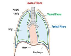
There are two pleural membranes surrounding the lung which naturally have a small amount of fluid between them this acts as a lubricant. The procedure allows your surgeon to take a biopsy of the pleura. This is a keyhole procedure involving two or three small incisions made between your ribs while you are under a general anaesthetic. The procedure allows your surgeon to see the inside of your chest using a flexible camera and for the passage of instruments to perform the procedure. Most patients following the procedure return to the ward with a chest drain in situ.
Why do I need a Video Assisted Pleural Biopsy
Your CT and or PET scan have shown an abnormality of your lung or pleura. The procedure would look to determine to cause of these changes. It may also be performed to drain fluid from around your lung with the aim of managing any future fluid building up, by performing a talc pleurodesis (instilling of talc between the lung and chest wall which acts as an irritant with the intention of fixing or sticking the lung to the chest wall) or the siting of a PleurX drain. A Pleurx drain drains fluid as it builds up or collects in the chest. At regular intervals a vacuumed bottle is attached to the tube to drain this fluid.
Can there be any complications or risks?
Potential risks of the procedure include chest or wound infection, bleeding, air leak or collapse of the lung.
How do I prepare for the biopsy
There is no specific preparation for the surgery however you should eat well and exercise within your limits, you should be as physically fit, and ell Nourished as this prepares you for surgery and your recovery afterwards. If you have signs of an infection for example a cough, cold or sore throat your procedure may be postponed.
What will happen?
- You will be admitted to ward 27 Castle Hill Hospital the day before your surgery
- Who will greeted by a member of the ward team on your arrival
- Your thoracic surgeon will perform your surgery
- The surgery will be performed under a general anaesthetic
- You will spend time in theatre recovery before returning to the ward following your surgery.
- Post-operative pain management will be offered by ward 27 nursing team
- Following your surgery, you will have a chest drain in situ, this will usually be removed before you go home.
What happens afterwards?
- You will be encouraged to be mobile following your surgery, you will feel sore and will require regular pain relief over the next couple of weeks
- Any biopsy results may take 7 to 10 days to process
- Recovery period is around 2 to 4 weeks
- Expected length of stay in hospital vary between 3 to 5 days
- You will be contacted by your surgeon two weeks following your discharge from the ward who will check on your recovery and explain the biopsy results. You may also be contacted by the Macmillan Thoracic Surgery Lung CNS Team with any further information for example if your case has been discussed in the Lung Cancer MDT.
- Follow-up treatment, this will depend on the biopsy findings
Should you require further advice on the issues contained in this leaflet, please do not hesitate to contact Ward 27 Castle Hill hospital on tel: 01482 461621 or the Macmillan Lung CNS Team on tel: 01482 461090.